techhub.social is one of the many independent Mastodon servers you can use to participate in the fediverse.
A hub primarily for passionate technologists, but everyone is welcome

Administered by:

Server stats:

4.6K
active users

#connectomics

0 posts0 participants0 posts today

The new Center for Structural and Functional #Connectomics (CSFC) will be established on our #Garching #campus with funding of around €69 million. 👏 Focus lies on the comprehensive mapping & analysis of all #neuronal connections in the brain: go.tum.de/373567

📷A.Eckert

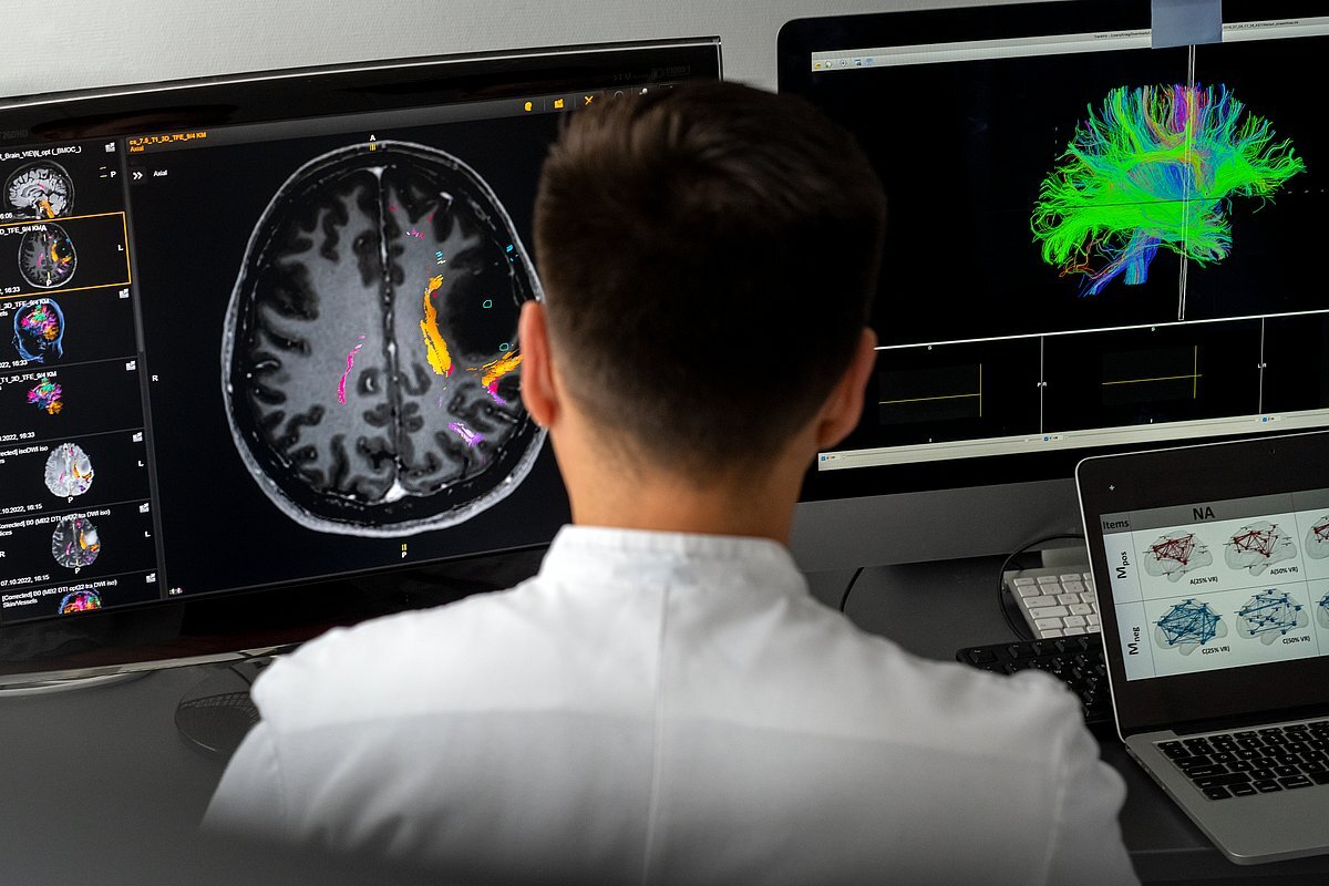
go.tum.deNew center for brain research on the Garching campusNew center for brain research: The TUM Center for Structural and Functional Connectomics will be established on the Garching campus.
Replied in thread

@biorxiv_neursci

#POINTseq : "(projections of interest by sequencing), a high-throughput and user-friendly barcoded connectomics method that uses cell type specific barcoding and sequencing to rapidly map single-cell projections of a cell type of interest for thousands of neurons per animal."

"We then applied POINTseq to midbrain dopaminergic neurons and reconstructed the brain-wide single-cell projections of 3,813 dopaminergic neurons in ventral tegmental area (VTA) and substantia nigra pars compacta (SNc)."

From Justus Kebschull's lab.

📰 "Cross-species comparative connectomics reveals the evolution of an olfactory circuit"
biorxiv.org/content/10.1101/20
#Neuroscience
#Connectomics
#Evolution
#Drosophila #Behaviour #Larva

bioRxiv · Cross-species comparative connectomics reveals the evolution of an olfactory circuitAnimal behavioural diversity ultimately stems from variation in neural circuitry, yet how central neural circuits evolve remains poorly understood. Studies of neural circuit evolution often focus on a few elements within a network. However, addressing fundamental questions in evolutionary neuroscience, such as whether some elements are more evolvable than others, requires a more global and unbiased approach. Here, we used synapse-level comparative connectomics to examine how an entire olfactory circuit evolves. We compared the full antennal lobe connectome of the larvae of two closely related Drosophila species, D. melanogaster and D. erecta, which differ in their ecological niches and odour-driven behaviours. We found that evolutionary change is unevenly distributed across the network. Some features, including neuron types, neuron numbers and interneuron-to-interneuron connectivity, are highly conserved. These conserved elements delineate a core circuit blueprint presumably required for fundamental olfactory processing. Superimposed on this scaffold, we find rewiring changes that mirror each species ecologies, including a systematic shift in the excitation-to-inhibition balance in the feedforward pathways. We further show that some neurons have changed more than others, and that even within individual neurons some synaptic elements remain conserved while others display major species-specific changes, suggesting evolutionary hot-spots within the circuit. Our findings reveal constrained and adaptable elements within olfactory networks, and establish a framework for identifying general principles in the evolution of neural circuits underlying behaviour. ### Competing Interest Statement C.S.X is the inventor of a US patent assigned to HHMI for the enhanced FIB-SEM systems used in this work: Xu, C.S., Hayworth K.J., Hess H.F. (2020) Enhanced FIB-SEM systems for large-volume 3D imaging. US Patent 10,600,615, 24 Mar 2020. European Molecular Biology Organization, ALTF1114-2024 International Human Frontier Science Program Organization, LT0036/2025-L, RGY0052/2022 European Research Council, 802531 Paul G. Allen Family Foundation Vallee Foundation, https://ror.org/05nmp3276 Chan Zuckerberg Initiative (United States), https://ror.org/02qenvm24, CP-2-1-Prieto-Godino The Francis Crick Institute, https://ror.org/04tnbqb63, CC2067, CC2240

From Elizabeth Marin at Zoology Dept., Cambridge University:

"Together with Greg Jefferis (MRC LMB, Cambridge), Wei-Chung Allen Lee (Harvard Medical School), and Meg Younger (Boston University), I have secured a £4.8M Wellcome Discovery Award to generate a mosquito brain connectome and investigate chemosensory circuits involved in human host-seeking."

"We are currently recruiting for two research assistant positions based in the Zoology department at Cambridge University. Please share this post with any likely candidates :)."

jobs.cam.ac.uk/job/51256/

www.jobs.cam.ac.ukResearch Assistant x 2 Connectomics Research Group (fixed term) - Job Opportunities - University of CambridgeResearch Assistant x 2 Connectomics Research Group (fixed term) in the Department of Zoology at the University of Cambridge.
Continued thread

This conundrum may originate in the cell type-centric view of connectivity, rather than considering the actual connectome into the analysis. The authors more or less say as much in the discussion:

"two cell types can have similar physiology and relatively similar connectivity without sharing input cell types. Overall, this analysis suggests that defining connection similarity by cell types may overly discretize the network, obscuring structure-function relationships."

"Infrequent strong connections constrain connectomic predictions of neuronal function", Currier and Clandinin
biorxiv.org/content/10.1101/20

Quite the reversal from studies showing that deriving connectomes from correlated neural activity is not accurate because of lacking a unique solution:

"we show that physiology is a stronger predictor of wiring than wiring is of physiology"

📰 "Serotonin selectively modulates visual responses of object motion detection in Drosophila"
biorxiv.org/content/10.1101/20
#Connectomics
#Drosophila

bioRxiv · Serotonin selectively modulates visual responses of object motion detection in DrosophilaSerotonin (5-HT) is a hormonal messenger that confers state-level changes upon the nervous system in both humans and flies. In Drosophila, lobula columnar (LC) cells are feature-detecting neurons that project from the optic lobe to the central brain, where each population forms an anatomically-distinct glomerulus with heterogeneous synaptic partners. Here, we investigated serotonin's effect on two LC populations with different 5-HT receptor expression profiles. Receptor expression does not predict neuromodulatory effects: LC15 expresses inhibitory 5-HT1A and 5-HT1B receptors, yet serotonin increases the amplitude of calcium responses to visual stimuli. LC12 expresses inhibitory 5-HT1A and excitatory 5-HT2A receptors, yet serotonin application does not influence visual responses. Serotonin targets select visual response properties, potentiating LC15 responses to a motion-defined bar and normalizing responses across bar velocity, but has no influence on contrast sensitivity. Serotonin does not significantly facilitate LC15 responses in postsynaptic dendrites, only in the presynaptic terminals of the glomerulus, which suggests that the neuromodulatory effects are strongest in the central brain. Connectomics confirms that LC12 and LC15 share neither presynaptic inputs nor postsynaptic outputs in the central brain. The wiring diagram shows no synaptic interactions between the LC15 circuit and major serotonergic 5-HTPLP neurons, nor to other serotonergic neurons of the central brain, suggesting that endogenous 5-HT acts via paracrine transmission on non-serotonergic pathways. Lobula- and glomerulus-specific GABAergic and glutamatergic inhibitory partners, positioned to filter visual stimuli, are putative 5-HT targets. These results provide a comparative framework for the neuromodulatory mechanisms involved in visual processing. ### Competing Interest Statement The authors have declared no competing interest.

A review: "C. elegans wired and wireless connectome: insights into principles of nervous system structure and function", by K Venkatesh, L Ripoll-Sánchez, I Beets, WR Schafer 2025
link.springer.com/article/10.1

SpringerLinkC. elegans wired and wireless connectome: insights into principles of nervous system structure and function - Journal of BiosciencesCaenorhabditis elegans is one of the primary model organisms for neuroscience research due to its well-annotated and compact nervous system. Being the first organism with a mapped connectome, published nearly 40 years ago, it holds a critical place in the field of neuroscience. Over the past decades, exhaustive mapping of the C. elegans nervous system at the molecular and cellular level, along with the development of tools to probe neural dynamics, have given invaluable insights on neuronal communication at the cellular, circuit, and systems level. In this review, we discuss key features of the C. elegans connectome, the wired (synaptic) as well as the wireless (extrasynaptic) network, and their role in executing complex behaviours. We delve into recent advances in C. elegans neuroscience, highlighting how in vivo and in silico studies have elucidated functional principles that govern sensory integration and the importance of assessing behavioural features at a systems level. With emerging connectomes of other, more complex organisms, this field offers a robust framework for testable hypotheses and comparative connectomics.

Now that's a big deal, and from a very credible source:

"Self-supervised image restoration in coherent X-ray neuronal microscopy", Laugros et al. (Alexandra Pacureanu) 2025
biorxiv.org/content/10.1101/20

"we present a self-supervised image restoration approach that simultaneously improves spatial resolution, contrast, and data acquisition speed. This enables revealing synapses with XNH, marking a major milestone in the quest for generating connectomes of full mammalian brains."

X-ray nanoholography took a turn towards higher resolution and higher throughput.

bioRxiv · Self-supervised image restoration in coherent X-ray neuronal microscopyCoherent X-ray microscopy is emerging as a transformative technology for neuronal imaging, with the potential to offer a scalable solution for reconstruction of neural circuits in millimeter sized tissue volumes. Specifically, X-ray holographic nanoto-mography (XNH) brings together outstanding capabilities in terms of contrast, spatial resolution and data acquisition speed. While recent XNH developments already enabled generating valuable datasets for neuro-sciences, a major challenge for reconstruction of neural circuits remained overcoming resolving power limits to distinguish smaller neurites and synapses in the reconstructed volumes. Here we present a self-supervised image restoration approach that simultaneously improves spatial resolution, contrast, and data acquisition speed. This enables revealing synapses with XNH, marking a major milestone in the quest for generating connectomes of full mammalian brains. We demonstrate that this method is effective for various types of neuronal tissues and acquisition schemes. We propose a scalable implementation compatible with multi-terabyte image volumes. Altogether, this work brings large-scale X-ray nanotomography to a new precision level. ### Competing Interest Statement The authors have declared no competing interest.
Replied in thread

@brembs This is very true as well in my field. Very incomplete data sets in #connectomics that then modelers pick up and run with, and don't understand when we show a lack of enthusiasm for their findings because the many limitations of the data weren't considered. To be fair, such limitations are as buried as possible in most manuscripts.

Replied in thread

@katchwreck For sure we won't understand how the brain works until the role of astrocytes and other glial cells is fully understood.
The #connectome though is understood as the wiring diagram where neurons are nodes and edges are synaptic connections. For additional interactions there's the "#neuromodulome" for e.g., neuropeptide/neuromodulator vs. the corresponding receptor, like in this paper by Lidia Ripoll-Sánchez et al. 2023 on C. elegans:
"The neuropeptidergic connectome of C. elegans" cell.com/neuron/fulltext/S0896
#neuroscience #Celegans #connectomics